Shingles Vaccine may be Viewed as a Quality of Life Vaccine

An example of shingles. (iStockphoto/Thinkstock)


A man with shingles of the face.

Shingles is caused by chickenpox virus called varicella zoster virus. The first indications that chickenpox and shingles were caused by the same virus were noticed at the beginning of the 20th century.

The incidence of shingles is mainly in adults. There are approximately four cases per 1000 population per year and a lifetime risk of 20 to 30 per cent.

Chickenpox generally occurs in children. Once the child gets over the illness the virus does not disappear from the body. Virus can settle down in one of the nerve cell bodies and lay dormant for many years.

When your resistance is low and this can be due to any reason, the virus may break out of the nerve cell and travel down the nerve causing viral infection of the skin in the area supplied by that nerve. This can happen decades after the chickenpox infection. Exactly how the virus remains latent in the body, and subsequently re-activates is not understood.

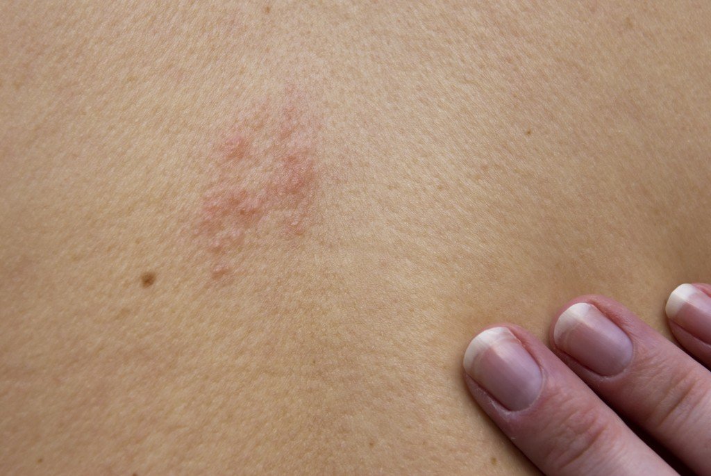
Shingles starts with burning pain, itching and tingling followed by painful rash and blisters in the area supplied by the affected nerve. The pain and rash most commonly occurs on the torso, but can appear on the face, eyes or other parts of the body. If the nerve to the eye is involved then a person may suffer loss of vision. It usually affects one nerve on one side of the body.

The rash and blisters heal within two to four weeks but some sufferers experience residual nerve pain for months or years. This condition is known as postherpetic neuralgia. About 20 per cent of patients with shingles suffer from this.

If the diagnosis of shingles is made early then it helps to start antiviral medications within 72 hours of the appearance of the rash. This reduces the severity and duration of the illness. The antiviral medications should be used for seven to ten days. The blisters crust over within seven to ten days, and usually the crusts fall off and the skin heals. But sometimes after severe blistering, scarring and discolored skin remains.

Until the rash has developed crusts, a person is extremely contagious. During the blister phase, direct contact with the rash can spread the virus to a person who has no immunity to the virus. This newly infected individual may then develop chickenpox, but will not immediately develop shingles.

Since 2008-2009, a vaccine for shingles is available for adults age 60 and over. The vaccine is used to boost the waning immunity to the virus that occurs with aging. The effectiveness of the vaccine is about 60 per cent. It is kind of a “quality of life” vaccine. It does not prevent death from shingles (an extremely rare event) but does help with postherpetic neuralgia (pain).

Booster doses of the vaccine are not recommended for healthy individuals. The efficacy of protection has not been assessed beyond four years and it is not known whether booster doses of vaccine are beneficial. This recommendation may need to be revisited as further information becomes available.

Start reading the preview of my book A Doctor's Journey for free on Amazon. Available on Kindle for $2.99!

Use of MRI in Evaluating Breast Lumps

A woman receiving a scan. (iStockphoto/Thinkstock)


A woman with locally advanced breast cancer.

The traditional way to assess a breast lump is to take a history, do a physical examination, do a fine needle aspiration cytology (examination of a breast lump aspirate under a microscope), mammogram and/or ultrasound, core biopsy under ultrasound control and finally, if there is no satisfactory answer then do a surgical biopsy.

A surgical biopsy gives us a definitive answer. But there are drawbacks to sending every patient with a breast lump for surgery. To start with it causes severe anxiety. You have to take a day off work. It requires local or general anaesthetic. There may or may not be postoperative complications like bleeding, bruising, discomfort, infection and pain.

On a long term basis, surgical biopsy will leave you with a scar and may be another lump which may be just a scar tissue but could be suspicious for cancer. Then you have to go through the whole process all over again.

Is there anything else we can do before going for surgery to make sure that there is no cancer in the breast?

You can ask for a second opinion. If all investigations are negative then there is a less than five per cent chance that cancer has been missed. In that case, we can leave the lump alone and provide follow up care with clinical examination and mammography or ultrasound, on a case by case basis. Sometimes a patient will ask for MRI.

MRI (magnetic resonance imaging) is a procedure that uses a magnet, radio waves, and a computer to make a series of detailed pictures of areas inside the body. MRI does not use any x-rays.
MRI is not available for routine screening. It is expensive and requires specialized equipment and personnel with good solid training to read the images. Hence, it is available in bigger cities only and is not covered by government insurance plans. MRI is more often used for breast imaging in the US than Canada because of the prevalence of private health care.

MRI is sensitive to small abnormalities in breast tissue. MRI also has limitations. For example, MRI cannot detect the presence of calcium deposits, which can be identified by mammography and may be a sign of cancer.

The value of breast MRI for breast cancer detection remains uncertain. And even at its best, MRI produces many uncertain findings. Some radiologists call these “unidentified bright objects,” or UBOs.

In women with a high inherited risk of breast cancer, screening trials of MRI breast scans have shown that MRI is more sensitive than mammography for finding breast tumors. Screening studies are ongoing.

Breast MRI is not recommended as a routine screening tool for breast cancer. However, for women at high risk, women with previous breast cancer, MRI can be useful in certain circumstances.

Start reading the preview of my book A Doctor's Journey for free on Amazon. Available on Kindle for $2.99!

Meckel’s Diverticulitis can Mimic Acute Appendicitis

A girl with lower abdominal pain. (iStockphoto/Thinkstock)


A specimen of acutely inflamed Meckel’s diverticulum.

It was nine o’clock in the evening. I was on-call for the general surgery group. After a busy day at the office and the hospital, and after a late supper, I had just sat down to watch some news on TV. The phone rings. My wife answers. She says to me, “It’s for you, honey. It’s the hospital emergency.”

The ER physician had just examined an ill looking seven-year old boy with right sided abdominal pain, nausea, vomiting and fever. The ER doctor wanted me to come and give a surgical opinion. The question I will be asked in ER is, “Does this boy have an acute appendicitis and does he need to go to OR for surgery this evening?”

After going through the boys history and physical examination, I came to the conclusion that the kid was quite sick with abdominal signs of acute appendicitis. Possibly perforated appendicitis and peritonitis. He was dehydrated. Intravenous fluids were given, preoperative antibiotics were given and he was taken to OR.

In the OR, as soon as the kid’s belly was opened, a large amount of purulent fluid poured out. The appendix looked normal. There was a hole in the small bowel where it meets the cecum (beginning of colon) where the appendix is located. The appendix, the terminal part of the small bowel and cecum were all stuck together due to the acute inflammation. To stop the leak from the small bowel, there was no choice but remove the terminal part of the small bowel, appendix and the cecum (called right hemicolectomy).

Postoperatively the child did very well. He went home nine days after surgery. Pathology of the specimen showed normal appendix, Meckel’s diverticulum with gastric mucosa with ulceration and perforation in the adjacent small bowel and peritonitis. Acid secretion from the gastric mucosa in the diverticulum had caused the ulceration and perforation.

A Meckel’s diverticulum is a true congenital diverticulum (bulge) in the small intestine present at birth. It is a vestigial remnant of the omphalomesenteric duct (also called the vitelline duct or yolk stalk), and is the most frequent malformation of the gastrointestinal tract.

It was first described by Fabricius Hildanus in the sixteenth century and later named after Johann Friedrich Meckel, who described the embryological origin of this type of diverticulum in 1809.

It is not that common. It is anti-mesenteric (on the free margin of the small bowel). For a medical student, the best memory aid is the rule of 2s: two per cent of the population, two feet from the ileocecal valve, two inches in length, two per cent are symptomatic, two types of common ectopic tissue (gastric and pancreatic), two years is the most common age at clinical presentation and two times more boys are affected.

Most people who are born with this have no symptoms. The most common presenting symptom is painless rectal bleeding such as black offensive stools, followed by intestinal obstruction, volvulus (torsion) and intussusception where a part of the intestine has invaginated into another section of intestine, similar to the way in which the parts of a collapsible telescope slide into one another. Over the years, I have seen examples of each one of the complication.

If a patient has symptoms and clinical diagnosis is not clear then it is worth doing a Meckel’s scan using technetium-99m (99mTc). This scan detects gastric mucosa; since approximately 50 per cent of symptomatic Meckel’s diverticula have ectopic gastric or pancreatic cells contained within them. Treatment is surgery.

Start reading the preview of my book A Doctor's Journey for free on Amazon. Available on Kindle for $2.99!

Gallstones can be a Source of Pain and Misery

Gallstones (iStockphoto/Thinkstock)

Clinical - Gall bladder and Gallstones
Left – gallbladder with stones. Right – 952 gallstones from my 35-year-old male patient who presented with chelecystitis, pancreatitis and common bile duct stones.

How many gallstones does it take to get symptoms?

Just one. The size and shape of the gallstone does not matter. The more stones you have the more risk of getting symptoms. There are many patients who have gallstones and have no symptoms. These patients do not require surgery until they are symptomatic or they are moving to a country where the health care system is not very reliable.

What is the world record for the number of gallstones removed from a single gallbladder?

According to a Medical Post (October 6, 1987) clipping I have on file, two British surgeons claimed a world record for the the number of gallstones removed from a single gallbladder – 23,530. This was in an 85-year-old woman who presented with severe colicky central abdominal pain. The gallstones are now in the pathology museum at St. Thomas Hospital medical school in London, England. I wonder who counted them all.

What is my record?

On October 8, 1987 I removed a gallbladder from a 35-year-old Saskatchewan man which contained 952 gallstones (see picture). I counted these myself! The man presented with gallstone pancreatitis and jaundice. He had severe upper abdominal pain and vomitting. He also had stones in the common bile duct. This is the duct which transports bile from the gallbladder to the intestine to help us digest food.

Who is the youngest patient to have gallbladder removed for gallstones?

According to Guinness World Records, an American child, Danylle Otteni was five years and six months old at the time she had her gallbladder and gallstones removed in Philadelphia, Pennsylvania, USA, on 14 October 2007. If I remember correctly, my youngest patient was around 14 years old.

Gallstones are very common in Western countries. Probably due to our diet which is high in refined and processed food. There are other factors which can contribute to gallstone formation. But we do not know exactly why some people have gallstones. There is some chemical imbalance in the bile which precipitates cholesterol to form a nucleus for a stone. On the basis of their composition, gallstones can be divided into the following types: cholesterol stones, pigment stones and mixed stones.

The treatment for symptomatic gallstones is surgery. It is the commonest elective general surgical procedure we do.

Once, the surgical procedure of choice was open cholecystectomy. In fact, Carl Langenbuch carried out the first cholecystectomy in 1882. Patients who undergo open cholecystectomy have a long surgical incision in the abdominal wall, have to stay in the hospital three to five days, consume fair amount of pain killers for post-operative pain and the recovery time at home is three to six weeks.

Things have changed in the last 15 to 20 years. Now most patients undergo laparoscopic cholecystectomy for symptomatic gallstones. Laparoscopic cholecystectomy was first performed in France in 1987. There are four tiny incisions in the abdominal wall, the hospital stay is usually overnight, the amount of pain killers required after surgery is minimal and the recovery time at home is usually less than one week. That is called progress.

Start reading the preview of my book A Doctor's Journey for free on Amazon. Available on Kindle for $2.99!