Photo Quiz: Scalp Lumps

Dermatofibrosarcoma Protuberans (Dr. Noorali Bharwani)

This is a 34-yr-old male with a history of scalp lumps. Is this a case of…
Lipoma?
Dermatofibrosarcoma? or
Sebaceous Cyst?

Correct Answer
Dermatofibrosarcoma Protuberans. This is a rare neoplasm of the dermis layer of the skin, and is classified as a sarcoma. It usually occurs in the trunk or extremities but can involve the head and neck particularly the scalp. In many respects, the disease behaves as a benign tumour, but in 2%-to-5% of cases it can metastasize, so it should be considered to have malignant potential.

This photo quiz was published on the Canadian Healthcare Network website.

Start reading the preview of my book A Doctor's Journey for free on Amazon. Available on Kindle for $2.99!

Merkel Cell Carcinoma

Left: Merkel cell carcinoma recurrence a few months after excision. Right: Progressive increase in size of recurrence over a few months. (Dr. Noorali Bharwani)

82 year-old male nursing home resident presented with a subcutaneous lump on the left knee for three to six months. Medical history includes head injury with intra-cranial bleed due to a fall, resulting in permanent physical disability and cognitive deficit.

The lump was about four cm in diameter. The patient and family requested excision biopsy as the patient kept drawing their attention to the lump that progressively turned red, as if it was getting inflamed. My first impression was that this was not a lipoma but probably an inflamed sebaceous cyst. It was excised under local anesthetic without any complications.

Pathology:

Merkel cell carcinoma of the skin, 4 cm in size. Peripheral and deep margins were extensively involved with lymphovascular invasion.


Merkel Cell Carcinoma (MCC)

Merkel cell carcinoma, a rare type of skin cancer, usually appears as a flesh to bluish-red colored nodule on sun-exposed areas, like the face, head and neck.

Merkel cell carcinoma is usually found in older people. Around 80% are caused by Merkel cell polyomavirus. Exposure to sunlight and a weak immune system increase the chance of developing MCC.

The tumor is locally invasive and also spreads rapidly through the body. Early diagnosis and treatment lowers the chance of metastasis.

Since the lesion has no distinguishable features from other skin cancers, the first treatment is surgical excision. Once the pathology report comes back the lesion can be identified as Merkel cell carcinoma. Further therapies like lymph node biopsy, radiation or chemotherapy can be considered if needed, to prevent metastasis and recurrence.


In this case, no further investigation or treatment were planned, as the patient’s quality of life would be extremely poor should he survive recurrence. The family decided it was time to let nature take its course.

Start reading the preview of my book A Doctor's Journey for free on Amazon. Available on Kindle for $2.99!

Parotid Gland Abscess

Abscess over the left parotid gland area. (Dr. Noorali Bharwani)

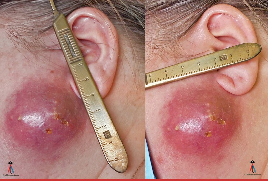
46 year-old female presented with a two-week history of an abscess over the left parotid area. The patient reports an abscess in the same location 18 months prior which she had aspirated. She has a medical history of Sjögren’s syndrome.

The patient had finished a course of amoxicillin-clavulanic acid and then was started on a course of a cephalosporin. Incision and drainage was done under local anesthetic and a large amount of pus was drained. The patient healed well.

Culture: Streptococcus anginosus

Streptococcus anginosus is part of the human bacteria flora, but can cause diseases including brain and liver abscesses under certain circumstances.” (Wikipedia)

Pathology: Showed no parotid tissue. There was acute-on-chronic inflammation of subcutaneous tissues.


Salivary Gland

Infection: parotitis, sialandenitis

Symptoms – One-sided salivary gland swelling. Fever and pain will accompany the swelling.

Pathogens – Typically normal bacteria found in the mouth. Viral infections such as mumps often affect the salivary glands. Mumps most often involves parotid salivary gland. Mumps is a rare problem today because of the MMR vaccine.

Dehydration and malnutrition raise the risk of getting a bacterial infection.

Sjögren’s syndrome

This is a chronic autoimmune disease in which cells of a person’s immune system attack the salivary and other moisture-producing glands, leading to dry mouth and eyes. About half of people with Sjögren’s syndrome also have enlargement of the salivary glands on both sides of the mouth, which is usually painless.

Treatment

In some cases, no treatment is needed.

Antibiotics for fever or pus drainage, or if the infection is caused by bacteria. Antibiotics are not useful against viral infections.

Surgery or aspiration to drain abscess.

Self-care steps that can be done at home to help with recovery:

  • Practice good oral hygiene. Brush teeth and floss well at least twice a day. This may help with healing and prevent infection from spreading.
  • Rinse mouth with warm salt-water rinses (1/2 teaspoon of salt in 1 cup of water) to ease pain keep the mouth moist.
  • Stop smoking.
  • Drink lots of water and use sugar-free lemon drops to increase the flow of saliva and reduce swelling.

Prognosis

Most salivary gland infections go away on or are cured with treatment. Some infections will return. Complications are uncommon.

Complications

  • Abscess of salivary gland
  • Infection returns
  • Spread of infection (cellulitis, Ludwig’s angina)

Prevention

In many cases, salivary gland infections cannot be prevented. Good oral hygiene may prevent some cases of bacterial infection.

Start reading the preview of my book A Doctor's Journey for free on Amazon. Available on Kindle for $2.99!

Pilomatricoma

Pilomatricoma before and after excision. (Dr. Noorali Bharwani)

A 47 year-old male presented with a three-month history of a lump below the right eyelid. It was slowly getting bigger. The lump was excised under local anaesthetic.

Pathology report

Specimen

Cyst right lower eyelid.
The specimen consists of a tan and brown irregularly shaped piece of skin measuring 1.3 x 0.5 x 0.2 cm. Attached is an underlying cyst-like structure measuring 1.0 x 0.5 x 0.5 cm. The cyst-like structure is extremely friable. The margins are marked with blue ink.

Diagnosis

Consistent with proliferating pilomatricoma


Pilomatricoma

Pilomatricoma is a benign cystic new growth.

Clinically, pilomatricoma usually presents in young individuals as a solitary cutaneous nodule with an average size of one cm and rarely exceeds 2 cm in diameter.

Proliferating pilomatricoma was first described in 1997.

The lesions are usually situated mostly on the head and neck.

The differential diagnosis includes classical pilomatricoma, pilomatrical carcinoma, and basal cell carcinoma.

These neoplasms should be excised with adequate surgical margins, and careful follow-up examinations are strongly recommended.

Proliferating pilomatricoma can occur at younger ages and should be considered in the differential diagnosis of solitary lesions in adults and children, even if the lesion is rapidly growing.

Start reading the preview of my book A Doctor's Journey for free on Amazon. Available on Kindle for $2.99!