Meckel’s Diverticulitis can Mimic Acute Appendicitis

A girl with lower abdominal pain. (iStockphoto/Thinkstock)


A specimen of acutely inflamed Meckel’s diverticulum.

It was nine o’clock in the evening. I was on-call for the general surgery group. After a busy day at the office and the hospital, and after a late supper, I had just sat down to watch some news on TV. The phone rings. My wife answers. She says to me, “It’s for you, honey. It’s the hospital emergency.”

The ER physician had just examined an ill looking seven-year old boy with right sided abdominal pain, nausea, vomiting and fever. The ER doctor wanted me to come and give a surgical opinion. The question I will be asked in ER is, “Does this boy have an acute appendicitis and does he need to go to OR for surgery this evening?”

After going through the boys history and physical examination, I came to the conclusion that the kid was quite sick with abdominal signs of acute appendicitis. Possibly perforated appendicitis and peritonitis. He was dehydrated. Intravenous fluids were given, preoperative antibiotics were given and he was taken to OR.

In the OR, as soon as the kid’s belly was opened, a large amount of purulent fluid poured out. The appendix looked normal. There was a hole in the small bowel where it meets the cecum (beginning of colon) where the appendix is located. The appendix, the terminal part of the small bowel and cecum were all stuck together due to the acute inflammation. To stop the leak from the small bowel, there was no choice but remove the terminal part of the small bowel, appendix and the cecum (called right hemicolectomy).

Postoperatively the child did very well. He went home nine days after surgery. Pathology of the specimen showed normal appendix, Meckel’s diverticulum with gastric mucosa with ulceration and perforation in the adjacent small bowel and peritonitis. Acid secretion from the gastric mucosa in the diverticulum had caused the ulceration and perforation.

A Meckel’s diverticulum is a true congenital diverticulum (bulge) in the small intestine present at birth. It is a vestigial remnant of the omphalomesenteric duct (also called the vitelline duct or yolk stalk), and is the most frequent malformation of the gastrointestinal tract.

It was first described by Fabricius Hildanus in the sixteenth century and later named after Johann Friedrich Meckel, who described the embryological origin of this type of diverticulum in 1809.

It is not that common. It is anti-mesenteric (on the free margin of the small bowel). For a medical student, the best memory aid is the rule of 2s: two per cent of the population, two feet from the ileocecal valve, two inches in length, two per cent are symptomatic, two types of common ectopic tissue (gastric and pancreatic), two years is the most common age at clinical presentation and two times more boys are affected.

Most people who are born with this have no symptoms. The most common presenting symptom is painless rectal bleeding such as black offensive stools, followed by intestinal obstruction, volvulus (torsion) and intussusception where a part of the intestine has invaginated into another section of intestine, similar to the way in which the parts of a collapsible telescope slide into one another. Over the years, I have seen examples of each one of the complication.

If a patient has symptoms and clinical diagnosis is not clear then it is worth doing a Meckel’s scan using technetium-99m (99mTc). This scan detects gastric mucosa; since approximately 50 per cent of symptomatic Meckel’s diverticula have ectopic gastric or pancreatic cells contained within them. Treatment is surgery.

Start reading the preview of my book A Doctor's Journey for free on Amazon. Available on Kindle for $2.99!

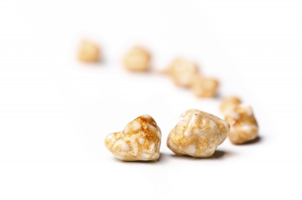
Gallstones can be a Source of Pain and Misery

Gallstones (iStockphoto/Thinkstock)

Clinical - Gall bladder and Gallstones
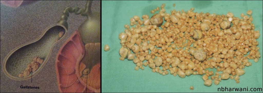
Left – gallbladder with stones. Right – 952 gallstones from my 35-year-old male patient who presented with chelecystitis, pancreatitis and common bile duct stones.

How many gallstones does it take to get symptoms?

Just one. The size and shape of the gallstone does not matter. The more stones you have the more risk of getting symptoms. There are many patients who have gallstones and have no symptoms. These patients do not require surgery until they are symptomatic or they are moving to a country where the health care system is not very reliable.

What is the world record for the number of gallstones removed from a single gallbladder?

According to a Medical Post (October 6, 1987) clipping I have on file, two British surgeons claimed a world record for the the number of gallstones removed from a single gallbladder – 23,530. This was in an 85-year-old woman who presented with severe colicky central abdominal pain. The gallstones are now in the pathology museum at St. Thomas Hospital medical school in London, England. I wonder who counted them all.

What is my record?

On October 8, 1987 I removed a gallbladder from a 35-year-old Saskatchewan man which contained 952 gallstones (see picture). I counted these myself! The man presented with gallstone pancreatitis and jaundice. He had severe upper abdominal pain and vomitting. He also had stones in the common bile duct. This is the duct which transports bile from the gallbladder to the intestine to help us digest food.

Who is the youngest patient to have gallbladder removed for gallstones?

According to Guinness World Records, an American child, Danylle Otteni was five years and six months old at the time she had her gallbladder and gallstones removed in Philadelphia, Pennsylvania, USA, on 14 October 2007. If I remember correctly, my youngest patient was around 14 years old.

Gallstones are very common in Western countries. Probably due to our diet which is high in refined and processed food. There are other factors which can contribute to gallstone formation. But we do not know exactly why some people have gallstones. There is some chemical imbalance in the bile which precipitates cholesterol to form a nucleus for a stone. On the basis of their composition, gallstones can be divided into the following types: cholesterol stones, pigment stones and mixed stones.

The treatment for symptomatic gallstones is surgery. It is the commonest elective general surgical procedure we do.

Once, the surgical procedure of choice was open cholecystectomy. In fact, Carl Langenbuch carried out the first cholecystectomy in 1882. Patients who undergo open cholecystectomy have a long surgical incision in the abdominal wall, have to stay in the hospital three to five days, consume fair amount of pain killers for post-operative pain and the recovery time at home is three to six weeks.

Things have changed in the last 15 to 20 years. Now most patients undergo laparoscopic cholecystectomy for symptomatic gallstones. Laparoscopic cholecystectomy was first performed in France in 1987. There are four tiny incisions in the abdominal wall, the hospital stay is usually overnight, the amount of pain killers required after surgery is minimal and the recovery time at home is usually less than one week. That is called progress.

Start reading the preview of my book A Doctor's Journey for free on Amazon. Available on Kindle for $2.99!
Surgery Is An Option For Individuals With Intractable Heartburn

A young man suffering from chest pain. (Hemera/Thinkstock)

Heartburn afflicts nearly two thirds of adults at some point in their lives, and accounts for millions of physician office visits every year. That adds up to a lot of tax dollars.

Last week we discussed the various aspects of medical treatment for heartburn, also known as gastroesophageal reflux disease (GERD).

There is a small group of patients with GERD who do not respond to medical treatment and they wonder if there is another option to relieve the symptoms and complications. Surgery, of course, is an option. Surgery is also an option for those who do respond to pills but cannot afford to buy them or do not want to be on the pills for the rest of their lives.

In GERD, there is failure of the antireflux barrier, allowing abnormal reflux of stomach contents into the esophagus. It is a mechanical disorder, which is caused by a defective lower esophageal sphincter (valve) at the junction of the esophagus and stomach, a gastric emptying disorder, or failed esophageal peristalsis. But the exact nature of the antireflux barrier is incompletely understood.

Surgery should not be considered without objective evidence of reflux. Based on the available evidence, the diagnosis of GERD can be confirmed if at least one of the following conditions exists: a mucosal break (inflammation and ulceration) seen on endoscopy in a patient with typical symptoms, Barrett’s esophagus on biopsy (considered to be a premalignant condition), a peptic stricture (narrowing due to chronic ulcer) in the absence of malignancy, or positive pH-metry (checks for the presence of acid reflux in the esophagus).

Clinical Guidelines published in 2010 by the Society of American Gastrointestinal and Endoscopic Surgeons (SAGES) makes the following recommendation regarding surgical management of GERD:

“Surgical therapy for GERD is an equally effective alternative to medical therapy and should be offered to appropriately selected patients by appropriately skilled surgeons. Surgical therapy effectively addresses the mechanical issues associated with the disease and results in long-term patient satisfaction. For surgery to compete with medical treatment, it has to be associated with minimal morbidity and cost.”

The antireflux surgery is done laparoscopically or by open method by surgeons thoroughly trained in this technique. Laparoscopic technique requires a short hospital stay and early return to work compared to open technique which requires a long abdominal incision and longer hospital stay with about six weeks of post op recovery. The two approaches have been demonstrated to have similar postoperative outcomes at the reported follow-up intervals (range three to 24 months) including reflux recurrence, dysphagia (difficulty swallowing), bloating, and reoperation rates.

SAGES recommendation says laparoscopic fundoplication should be preferred over its open alternative as it is associated with superior early outcomes (shorter hospital stay and return to normal activities, and fewer complications) and no significant differences in late outcomes (failure rates). If you are referred to a surgeon for this procedure then you should carefully discuss pros and cons of medical vs. surgical treatment. Your case may be different than your neighbour’s. What is good for your neighbour may not be good for you.

Start reading the preview of my book A Doctor's Journey for free on Amazon. Available on Kindle for $2.99!