Use of MRI in Evaluating Breast Lumps

A woman receiving a scan. (iStockphoto/Thinkstock)


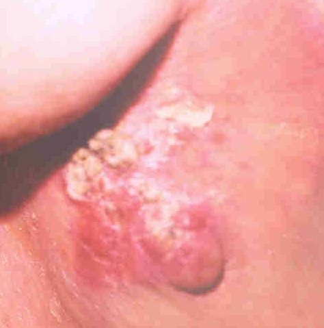
A woman with locally advanced breast cancer.

The traditional way to assess a breast lump is to take a history, do a physical examination, do a fine needle aspiration cytology (examination of a breast lump aspirate under a microscope), mammogram and/or ultrasound, core biopsy under ultrasound control and finally, if there is no satisfactory answer then do a surgical biopsy.

A surgical biopsy gives us a definitive answer. But there are drawbacks to sending every patient with a breast lump for surgery. To start with it causes severe anxiety. You have to take a day off work. It requires local or general anaesthetic. There may or may not be postoperative complications like bleeding, bruising, discomfort, infection and pain.

On a long term basis, surgical biopsy will leave you with a scar and may be another lump which may be just a scar tissue but could be suspicious for cancer. Then you have to go through the whole process all over again.

Is there anything else we can do before going for surgery to make sure that there is no cancer in the breast?

You can ask for a second opinion. If all investigations are negative then there is a less than five per cent chance that cancer has been missed. In that case, we can leave the lump alone and provide follow up care with clinical examination and mammography or ultrasound, on a case by case basis. Sometimes a patient will ask for MRI.

MRI (magnetic resonance imaging) is a procedure that uses a magnet, radio waves, and a computer to make a series of detailed pictures of areas inside the body. MRI does not use any x-rays.
MRI is not available for routine screening. It is expensive and requires specialized equipment and personnel with good solid training to read the images. Hence, it is available in bigger cities only and is not covered by government insurance plans. MRI is more often used for breast imaging in the US than Canada because of the prevalence of private health care.

MRI is sensitive to small abnormalities in breast tissue. MRI also has limitations. For example, MRI cannot detect the presence of calcium deposits, which can be identified by mammography and may be a sign of cancer.

The value of breast MRI for breast cancer detection remains uncertain. And even at its best, MRI produces many uncertain findings. Some radiologists call these “unidentified bright objects,” or UBOs.

In women with a high inherited risk of breast cancer, screening trials of MRI breast scans have shown that MRI is more sensitive than mammography for finding breast tumors. Screening studies are ongoing.

Breast MRI is not recommended as a routine screening tool for breast cancer. However, for women at high risk, women with previous breast cancer, MRI can be useful in certain circumstances.

Start reading the preview of my book A Doctor's Journey for free on Amazon. Available on Kindle for $2.99!

Meckel’s Diverticulitis can Mimic Acute Appendicitis

A girl with lower abdominal pain. (iStockphoto/Thinkstock)


A specimen of acutely inflamed Meckel’s diverticulum.

It was nine o’clock in the evening. I was on-call for the general surgery group. After a busy day at the office and the hospital, and after a late supper, I had just sat down to watch some news on TV. The phone rings. My wife answers. She says to me, “It’s for you, honey. It’s the hospital emergency.”

The ER physician had just examined an ill looking seven-year old boy with right sided abdominal pain, nausea, vomiting and fever. The ER doctor wanted me to come and give a surgical opinion. The question I will be asked in ER is, “Does this boy have an acute appendicitis and does he need to go to OR for surgery this evening?”

After going through the boys history and physical examination, I came to the conclusion that the kid was quite sick with abdominal signs of acute appendicitis. Possibly perforated appendicitis and peritonitis. He was dehydrated. Intravenous fluids were given, preoperative antibiotics were given and he was taken to OR.

In the OR, as soon as the kid’s belly was opened, a large amount of purulent fluid poured out. The appendix looked normal. There was a hole in the small bowel where it meets the cecum (beginning of colon) where the appendix is located. The appendix, the terminal part of the small bowel and cecum were all stuck together due to the acute inflammation. To stop the leak from the small bowel, there was no choice but remove the terminal part of the small bowel, appendix and the cecum (called right hemicolectomy).

Postoperatively the child did very well. He went home nine days after surgery. Pathology of the specimen showed normal appendix, Meckel’s diverticulum with gastric mucosa with ulceration and perforation in the adjacent small bowel and peritonitis. Acid secretion from the gastric mucosa in the diverticulum had caused the ulceration and perforation.

A Meckel’s diverticulum is a true congenital diverticulum (bulge) in the small intestine present at birth. It is a vestigial remnant of the omphalomesenteric duct (also called the vitelline duct or yolk stalk), and is the most frequent malformation of the gastrointestinal tract.

It was first described by Fabricius Hildanus in the sixteenth century and later named after Johann Friedrich Meckel, who described the embryological origin of this type of diverticulum in 1809.

It is not that common. It is anti-mesenteric (on the free margin of the small bowel). For a medical student, the best memory aid is the rule of 2s: two per cent of the population, two feet from the ileocecal valve, two inches in length, two per cent are symptomatic, two types of common ectopic tissue (gastric and pancreatic), two years is the most common age at clinical presentation and two times more boys are affected.

Most people who are born with this have no symptoms. The most common presenting symptom is painless rectal bleeding such as black offensive stools, followed by intestinal obstruction, volvulus (torsion) and intussusception where a part of the intestine has invaginated into another section of intestine, similar to the way in which the parts of a collapsible telescope slide into one another. Over the years, I have seen examples of each one of the complication.

If a patient has symptoms and clinical diagnosis is not clear then it is worth doing a Meckel’s scan using technetium-99m (99mTc). This scan detects gastric mucosa; since approximately 50 per cent of symptomatic Meckel’s diverticula have ectopic gastric or pancreatic cells contained within them. Treatment is surgery.

Start reading the preview of my book A Doctor's Journey for free on Amazon. Available on Kindle for $2.99!

Gallstones can be a Source of Pain and Misery

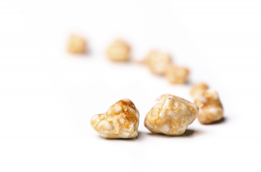
Gallstones (iStockphoto/Thinkstock)

Clinical - Gall bladder and Gallstones
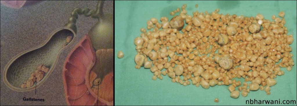
Left – gallbladder with stones. Right – 952 gallstones from my 35-year-old male patient who presented with chelecystitis, pancreatitis and common bile duct stones.

How many gallstones does it take to get symptoms?

Just one. The size and shape of the gallstone does not matter. The more stones you have the more risk of getting symptoms. There are many patients who have gallstones and have no symptoms. These patients do not require surgery until they are symptomatic or they are moving to a country where the health care system is not very reliable.

What is the world record for the number of gallstones removed from a single gallbladder?

According to a Medical Post (October 6, 1987) clipping I have on file, two British surgeons claimed a world record for the the number of gallstones removed from a single gallbladder – 23,530. This was in an 85-year-old woman who presented with severe colicky central abdominal pain. The gallstones are now in the pathology museum at St. Thomas Hospital medical school in London, England. I wonder who counted them all.

What is my record?

On October 8, 1987 I removed a gallbladder from a 35-year-old Saskatchewan man which contained 952 gallstones (see picture). I counted these myself! The man presented with gallstone pancreatitis and jaundice. He had severe upper abdominal pain and vomitting. He also had stones in the common bile duct. This is the duct which transports bile from the gallbladder to the intestine to help us digest food.

Who is the youngest patient to have gallbladder removed for gallstones?

According to Guinness World Records, an American child, Danylle Otteni was five years and six months old at the time she had her gallbladder and gallstones removed in Philadelphia, Pennsylvania, USA, on 14 October 2007. If I remember correctly, my youngest patient was around 14 years old.

Gallstones are very common in Western countries. Probably due to our diet which is high in refined and processed food. There are other factors which can contribute to gallstone formation. But we do not know exactly why some people have gallstones. There is some chemical imbalance in the bile which precipitates cholesterol to form a nucleus for a stone. On the basis of their composition, gallstones can be divided into the following types: cholesterol stones, pigment stones and mixed stones.

The treatment for symptomatic gallstones is surgery. It is the commonest elective general surgical procedure we do.

Once, the surgical procedure of choice was open cholecystectomy. In fact, Carl Langenbuch carried out the first cholecystectomy in 1882. Patients who undergo open cholecystectomy have a long surgical incision in the abdominal wall, have to stay in the hospital three to five days, consume fair amount of pain killers for post-operative pain and the recovery time at home is three to six weeks.

Things have changed in the last 15 to 20 years. Now most patients undergo laparoscopic cholecystectomy for symptomatic gallstones. Laparoscopic cholecystectomy was first performed in France in 1987. There are four tiny incisions in the abdominal wall, the hospital stay is usually overnight, the amount of pain killers required after surgery is minimal and the recovery time at home is usually less than one week. That is called progress.

Start reading the preview of my book A Doctor's Journey for free on Amazon. Available on Kindle for $2.99!
Bob Marley and Importance of Melanoma in Darker-Pigmented Population

A reggae hat. (iStockphoto/Thinkstock)


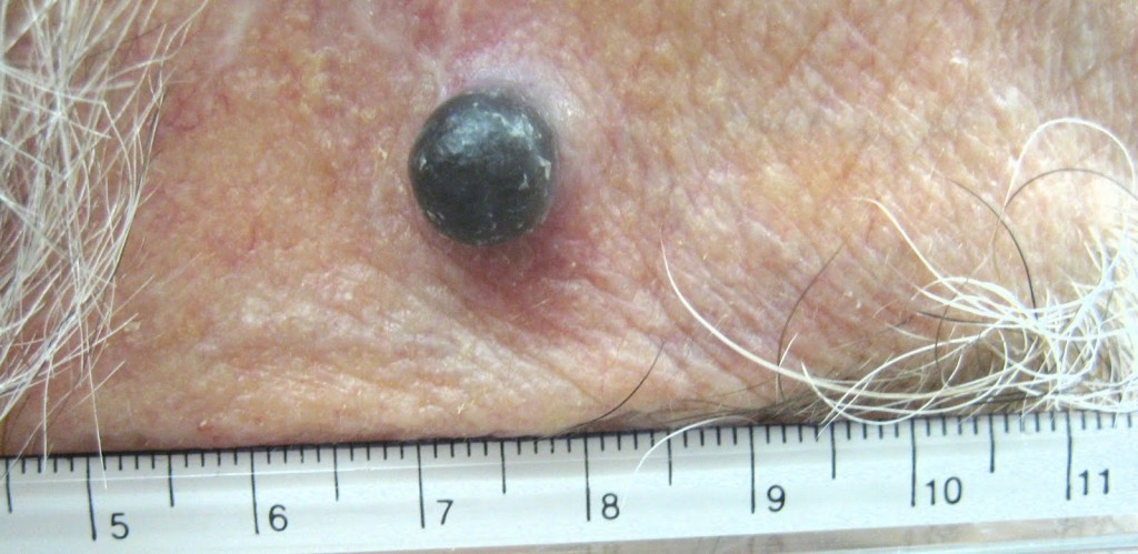
A case of recurrent melanoma in a Caucasian male – temple area.

I enjoy Bob Marley music. I listen to his songs quite often. As most of you know, Bob Marley was a Jamaican singer-songwriter and musician. In May 1981, he died at a very young age of 36 from melanoma. This was tragic. If he would have listened to his doctors then, who knows, he would still be around entertaining us with live performances.

In 1977, Marley was found to have malignant melanoma under the nail of one of his toes. Marley turned down doctors’ advice to have his toe amputated, citing his religious beliefs. He followed the Rastafari tradition. The spread of melanoma to his lungs and brain caused his death. Before his death, he is reported to have said, “Money can’t buy life”.

Bob Marley’s case is interesting from a medical point of view. First, melanoma is not that common in black population and secondly, melanoma under the nail (subungual) is not common either. It is an accepted fact that malignant melanoma in black population and other minority ethnic populations represents an aggressive disease highly associated with invasive lesions. They present with more advanced stage of disease at diagnosis, and consequently with a decreased survival compared with Caucasians.

Melanoma is the sixth most common cancer in North America and the single most common one among young adults 25-29 years old. Lifetime risk of developing melanoma in whites is currently estimated at 1 in 50, compared to 1 in 1000 in African-Americans.

Darker-pigmented populations are consistently reported to have lower risk for melanoma, possibly related to protection from ultraviolet radiation (UVR) provided by melanin.

Melanin is the primary determinant of skin color. It is also found in hair, the pigmented tissue underlying the iris of the eye, and other pigmented areas of the body and brain. The melanin in the skin is produced by cells called melanocytes. Some individuals have very little or no melanin in their bodies, a condition known as albinism.

Production of melanin is stimulated by DNA damage induced by UVB-radiation, and it leads to a delayed development of a tan. It is an excellent photoprotectant. This is because it efficiently absorbs harmful UV-radiation (ultraviolet) and transforms the energy into harmless heat. This prevents the indirect DNA damage that is responsible for the formation of malignant melanoma and other skin cancers.

Caucasians have a predilection to develop lesions on sun-exposed surfaces, including face and neck. Blacks have lesions predominantly located on sun-protected mucosal and acral sites.

Acral sites are the palms, soles, under the nails and in the mouth. It occurs on non hair-bearing surfaces of the body which may or may not be exposed to sunlight. Unlike other forms of melanoma, acral lentiginous melanoma (ALM) does not appear to be linked to sun exposure.

Lentiginous means small, flat, pigmented spot on the skin or under the nail. The reason these lesions have poor prognosis is because they are quite often clinically misdiagnosed.

In fact, an estimated one-third to one-half of all cases of ALM are incorrectly diagnosed at initial presentation as the more commonly appearing benign skin lesions including warts, infections, ulcers, callus, traumatic wounds, and blood clots. Some melanomas have no pigment and these are hard to diagnose early unless you notice some change.

The moral of today’s story is: be vigilant, protect against UV rays, and report to your doctor if there is any change in a mole. If you have a pigmented lesion under a nail, palm of your hands, or sole of your feet which does not go away (a blood clot will slowly disappear) then get a biopsy done. And listen to your doctor. Melanoma can be cured if picked up early.

Long live Bob Marley and his music.

Start reading the preview of my book A Doctor's Journey for free on Amazon. Available on Kindle for $2.99!