Use of MRI in Evaluating Breast Lumps

A woman receiving a scan. (iStockphoto/Thinkstock)


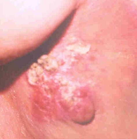
A woman with locally advanced breast cancer.

The traditional way to assess a breast lump is to take a history, do a physical examination, do a fine needle aspiration cytology (examination of a breast lump aspirate under a microscope), mammogram and/or ultrasound, core biopsy under ultrasound control and finally, if there is no satisfactory answer then do a surgical biopsy.

A surgical biopsy gives us a definitive answer. But there are drawbacks to sending every patient with a breast lump for surgery. To start with it causes severe anxiety. You have to take a day off work. It requires local or general anaesthetic. There may or may not be postoperative complications like bleeding, bruising, discomfort, infection and pain.

On a long term basis, surgical biopsy will leave you with a scar and may be another lump which may be just a scar tissue but could be suspicious for cancer. Then you have to go through the whole process all over again.

Is there anything else we can do before going for surgery to make sure that there is no cancer in the breast?

You can ask for a second opinion. If all investigations are negative then there is a less than five per cent chance that cancer has been missed. In that case, we can leave the lump alone and provide follow up care with clinical examination and mammography or ultrasound, on a case by case basis. Sometimes a patient will ask for MRI.

MRI (magnetic resonance imaging) is a procedure that uses a magnet, radio waves, and a computer to make a series of detailed pictures of areas inside the body. MRI does not use any x-rays.
MRI is not available for routine screening. It is expensive and requires specialized equipment and personnel with good solid training to read the images. Hence, it is available in bigger cities only and is not covered by government insurance plans. MRI is more often used for breast imaging in the US than Canada because of the prevalence of private health care.

MRI is sensitive to small abnormalities in breast tissue. MRI also has limitations. For example, MRI cannot detect the presence of calcium deposits, which can be identified by mammography and may be a sign of cancer.

The value of breast MRI for breast cancer detection remains uncertain. And even at its best, MRI produces many uncertain findings. Some radiologists call these “unidentified bright objects,” or UBOs.

In women with a high inherited risk of breast cancer, screening trials of MRI breast scans have shown that MRI is more sensitive than mammography for finding breast tumors. Screening studies are ongoing.

Breast MRI is not recommended as a routine screening tool for breast cancer. However, for women at high risk, women with previous breast cancer, MRI can be useful in certain circumstances.

Start reading the preview of my book A Doctor's Journey for free on Amazon. Available on Kindle for $2.99!

Skin Blemishes of Breast

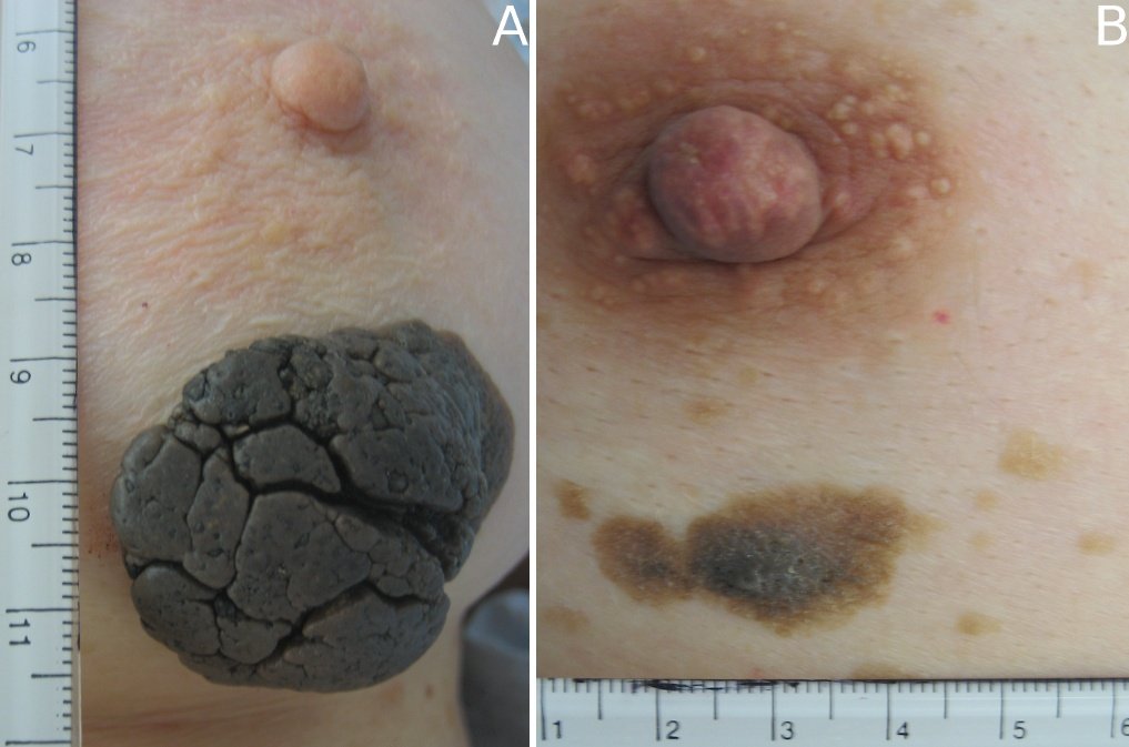
Skin Blemish of Breast
(click to enlarge)

Skin lesions and blemishes are very common. Most of them are benign and have no malignant potential. But some can be malignant or potentially malignant. Some areas of the body are easy to examine but other parts of the body are not clearly visible. One such area is lower part of a woman’s breast.

Many women are very particular in doing breast self-examination but forget to do visual inspection of the nipples, areola and under surface of the breasts where moles can be missed.

Moles that are of medical concern are those that look different than other existing moles or those that first appear after age 20. If you notice changes in a mole’s colour, height, size or shape, you should have these moles checked. If the moles bleed, ooze, itch, appear scaly or become tender or painful then it is time to have them removed and checked for cancer.

The following ABCDEs are important signs of moles that could be cancerous:

Asymmetry – one half of the mole does not match the other half.

Border – the border or edges of the mole are ragged, blurred or irregular.

Colour – the colour of the mole is not the same throughout or has shades of tan, brown, black, blue, white or red.

Diameter – the diameter of a mole is six millimetres or larger.

Evolution – moles which have changed over a period of time.

Remember, there is a forecast for a sizzling summer this year. So, we should continue to remind ourselves to protect the rest of the body from sunburn. Use of sunscreen is one way to do it. There is a lot of misconception about what kind of sunscreen to use and how to apply.

Sun Protection Factor (SPF) 30 provides 30 times greater sun protection than unprotected skin when exposed to damaging sun’s ultraviolet B (UVB) rays. This does not mean you can stay in the sun for 30 hours without burning yourself. Sunscreen should be applied liberally and often depending on how much you sweat and how wet you are. Make sure the sunscreen blocks UVB and UVA. Higher SPF provides better protection. Wear protective clothings, wide brimmed hat, sunglasses which block both ultraviolet rays.

The best protection against sun’s damaging rays is to stay away from the sun and take your vitamin D regularly. I guess that is too much to ask, especially when our summers are so short. So enjoy the sun but be sun smart.

Start reading the preview of my book A Doctor's Journey for free on Amazon. Available on Kindle for $2.99!

New Guidelines for Hormone Replacement Therapy

In 2002, we learnt that in post-menopausal women, hormone replacement therapy (HRT) with estrogen was doing more harm than good. The study, Women’s Health Initiative trial, involved 16,608 post-menopausal women, aged 50 to 79. Because of these findings, the study was discontinued early.

The harmful effects of estrogen therapy were: 41 percent increase in stroke, 29 percent increase in heart attacks, doubling of rates of blood clots in the legs and lungs, 26 percent increase in breast cancer and 22 percent increase in total cardiovascular disease.

But the report also said HRT has benefits: 37 percent reduction in cases of colorectal cancer, 33 percent reduction in hip fractures, no difference in total death rate from all causes and controls hot flashes.

Women have been taking estrogen to relieve post-menopausal symptoms for many years. In 1940s, pharmaceutical companies started producing estrogen from pregnant mare’s urine called Premarin. Twenty years later, the drug was being recommended for women who showed evidence of estrogen lack. Practically, all women over the age of 50. But things changed after 2002. According media reports, the highly publicized research led to a sharp drop in HRT prescriptions, to about five million prescriptions last year from 12 million in 2002.

The new guidelines from the Society of Obstetricians and Gynaecologists of Canada say hormone replacement therapy is safe and effective when used immediately at the onset of menopause and for a relatively short time. That means it would be safe to use HRT in women in their 50s to relieve their hot flashes, night sweats and vaginal dryness. And they should discontinue using HRT within five years.

The new guidelines were announced after a committee of experts re-examined the data from the previous study and came to the conclusion that the age at which women begin taking HRT, the dose and the duration all have an influence on risk. The guidelines do not endorse the use of complementary therapies as there is little or no evidence that herbal products sold for the treatment of menopausal symptoms have any benefit.

Women are advised to take control of their lives. Many problems (mood swings, insomnia, osteoporosis and difficulty concentrating) can be alleviated with lifestyle changes – weight control, healthy eating, exercise, no smoking, stress relief and meditation.

So, what should women do now? Why do experts change their minds about such things? First they create a panic then it takes them seven years to come back and say you are ok to take it within certain limitations.

I guess that is the nature of medical science. There is so much we know and again there is so much we do not know. Physicians find this as frustrating as the general public. Some women are going to be quite upset, may be even angry and frustrated, that they were deprived of HRT when they needed it. But it is never too late to sit down with your doctor and discuss your symptoms and indication for HRT.

Start reading the preview of my book A Doctor's Journey for free on Amazon. Available on Kindle for $2.99!

Breast Lumps Should Not Be Ignored

Breast lumps are very common. Breast lumps appear in women and men. They may appear in children. But mostly the breast lumps appear in adults. A breast lump is always an important clinical finding – never to be ignored. It is also a source of anxiety for the patient and his or her family. The first thing they want to know is, “Doctor, is this cancer?”

Investigations of a breast lump starts with a history and physical examination. Physical examination is best done between seven to 10 days from the first day of the menstrual cycle. The clinical history will establish how long the lump has been present. Whether there has been any change. And if there is a previous history of breast biopsy or breast cancer.

Risk factors for breast cancer will be noted (patient’s age and family history). It is important to remember, absence of risk factors does not decrease the probability of cancer.

Careful examination of the breasts will confirm the presence and establish the character of the lump. Hard, irregular, tethered, fixed or painless lump may be suggestive of cancer. Axillary (armpit) and neck areas are checked for lymph glands. The predictive value of physical examination (to say whether the lump is benign or malignant) in experienced hands is about 75 percent.

Mammograms in younger women are not very helpful. But after mid-30s the value of mammography increases. Quite often mammography can clarify the nature of the lump. It can also detect non-palpable lumps.

The overall level of sensitivity of mammography in palpable breast cancers may be no more than 82 percent. It may be even lower in premenopausal women. A negative mammogram in the presence of a persistent lump does not exclude malignancy.

Fine-needle aspiration biopsy done in the office can establish whether a breast lump is solid or cystic. When the lump is solid, cells can be aspirated for examination under a microscope. If fluid is obtained during aspiration and the lump disappears then it is a cyst.

In a cystic lump, if the fluid is not bloody then no specimen needs to be sent for examination and no further investigation is necessary. Bloody fluid may be suggestive of cancer and should be sent for microscopic examination.

Fine-needle aspiration biopsy should provide satisfactory specimen for microscopic examination in 90 percent of the cases with an accuracy rate of 95 percent to detect presence of cancer cells.

When physical examination, mammography, and microscopic examination of aspirated specimen indicate cancer then the probability of this being confirmed by surgery is more than 99 percent. If all three tests are negative then the lump being cancerous is less than one percent.

Core-biopsy, where a bigger sample (a core of tissue) is taken from a breast lump, has 90 percent predictive value for breast cancer. A negative biopsy may make it unnecessary to do an open surgical biopsy.

Ultrasound of the breast lump is an alternative method to distinguish a cyst from a solid lump. Ultrasound is useful when a non-palpable lump is seen on a mammogram.

Nuclear medicine techniques (Miraluma scan) have been used to evaluate breast cancer. Its predictive value for cancer is around 80 percent for a palpable lesion and 50 to 70 percent for a non-palpable breast lump found on mammography.

Whenever reasonable doubt remains about cancer (since none of the tests have a one hundred percent accuracy rate), a surgical open biopsy is undertaken. Only about one in every five to 10 breast biopsies leads to a diagnosis of cancer. But every breast lump should be investigated thoroughly.

Start reading the preview of my book A Doctor's Journey for free on Amazon. Available on Kindle for $2.99!