Inflammatory Breast Cancer is a Rare and Very Aggressive Disease

A woman making a heart frame for the pink badge. (iStockphoto/Thinkstock)

Inflammatory breast cancer accounts for less than five percent of all breast cancers diagnosed in North America. Most inflammatory breast cancers are invasive ductal carcinomas. They develop from cells that line the milk ducts of the breast and then spread beyond the ducts.

It is a very aggressive disease with symptoms that include redness, swelling, tenderness, and warmth in the breast. As if you have an abscess of the breast. But it may be cancer.

The breast swells up because cancer cells block lymph vessels in the skin of the breast. The disease progresses rapidly, often in a matter of weeks or months. By the time the diagnosis is made it is either stage III or IV, depending on whether cancer cells have spread only to nearby lymph nodes (stage III) or to other tissues as well (stage IV).

It is more common and diagnosed at younger ages (median age of 57 years, compared with a median age of 62 years for other types of breast cancer). It is more common in African American women than in white women. It is more common in obese women than in women of normal weight. It can occur in men.

Rapid diagnosis and treatment is key to successful treatment. An international panel of experts published guidelines on how doctors can diagnose and stage inflammatory breast cancer correctly. These are:

1. A rapid onset of redness, swelling, and a peau d’orange (skin of an orange) appearance and/or abnormal breast warmth, with or without a lump that can be felt.

2. The above-mentioned symptoms have been present for less than 6 months.

3. The redness covers at least a third of the breast.

4. Initial biopsy samples from the affected breast show invasive carcinoma.

A diagnostic mammogram and an ultrasound of the breast and regional (nearby) lymph nodes is part of the diagnostic workup. A PET scan or a CT scan and a bone scan is done to see if the cancer has spread to other parts of the body.

Inflammatory breast cancer is treated first with systemic chemotherapy to help shrink the tumor, then with surgery to remove the tumor, followed by radiation therapy. This approach to treatment is called a multimodal approach. Studies have found that women with inflammatory breast cancer who are treated with a multi-modal approach have better responses to therapy and longer survival. If a woman’s biopsy samples show that her cancer cells contain hormone receptors, hormone therapy is another treatment option.

What is the prognosis of patients with inflammatory breast cancer?

Since this is an aggressive tumour, in general, women with inflammatory breast cancer do not survive as long as women diagnosed with other types of breast cancer. According to statistics from National Cancer Institute’s Surveillance, Epidemiology, and End Results (SEER) program, the 5-year relative survival for women diagnosed with inflammatory breast cancer during the period from 1988 through 2001 was 34 percent, compared with a 5-year relative survival of up to 87 percent among women diagnosed with other stages of invasive breast cancers.

National Cancer Institute’s website encourages women with inflammatory breast cancer to voluntary for ongoing research. The research, especially at the molecular level, will increase our understanding of how inflammatory breast cancer begins and progresses. This knowledge should enable the development of new treatments and more accurate prognoses for women diagnosed with this disease. It is important, therefore, that women who are diagnosed with inflammatory breast cancer talk with their doctor about the option of participating in a clinical trial.

Start reading the preview of my book A Doctor's Journey for free on Amazon. Available on Kindle for $2.99!

Bob Marley and Importance of Melanoma in Darker-Pigmented Population

A reggae hat. (iStockphoto/Thinkstock)


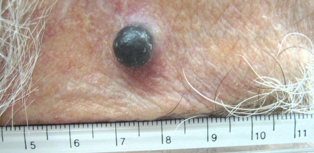
A case of recurrent melanoma in a Caucasian male – temple area.

I enjoy Bob Marley music. I listen to his songs quite often. As most of you know, Bob Marley was a Jamaican singer-songwriter and musician. In May 1981, he died at a very young age of 36 from melanoma. This was tragic. If he would have listened to his doctors then, who knows, he would still be around entertaining us with live performances.

In 1977, Marley was found to have malignant melanoma under the nail of one of his toes. Marley turned down doctors’ advice to have his toe amputated, citing his religious beliefs. He followed the Rastafari tradition. The spread of melanoma to his lungs and brain caused his death. Before his death, he is reported to have said, “Money can’t buy life”.

Bob Marley’s case is interesting from a medical point of view. First, melanoma is not that common in black population and secondly, melanoma under the nail (subungual) is not common either. It is an accepted fact that malignant melanoma in black population and other minority ethnic populations represents an aggressive disease highly associated with invasive lesions. They present with more advanced stage of disease at diagnosis, and consequently with a decreased survival compared with Caucasians.

Melanoma is the sixth most common cancer in North America and the single most common one among young adults 25-29 years old. Lifetime risk of developing melanoma in whites is currently estimated at 1 in 50, compared to 1 in 1000 in African-Americans.

Darker-pigmented populations are consistently reported to have lower risk for melanoma, possibly related to protection from ultraviolet radiation (UVR) provided by melanin.

Melanin is the primary determinant of skin color. It is also found in hair, the pigmented tissue underlying the iris of the eye, and other pigmented areas of the body and brain. The melanin in the skin is produced by cells called melanocytes. Some individuals have very little or no melanin in their bodies, a condition known as albinism.

Production of melanin is stimulated by DNA damage induced by UVB-radiation, and it leads to a delayed development of a tan. It is an excellent photoprotectant. This is because it efficiently absorbs harmful UV-radiation (ultraviolet) and transforms the energy into harmless heat. This prevents the indirect DNA damage that is responsible for the formation of malignant melanoma and other skin cancers.

Caucasians have a predilection to develop lesions on sun-exposed surfaces, including face and neck. Blacks have lesions predominantly located on sun-protected mucosal and acral sites.

Acral sites are the palms, soles, under the nails and in the mouth. It occurs on non hair-bearing surfaces of the body which may or may not be exposed to sunlight. Unlike other forms of melanoma, acral lentiginous melanoma (ALM) does not appear to be linked to sun exposure.

Lentiginous means small, flat, pigmented spot on the skin or under the nail. The reason these lesions have poor prognosis is because they are quite often clinically misdiagnosed.

In fact, an estimated one-third to one-half of all cases of ALM are incorrectly diagnosed at initial presentation as the more commonly appearing benign skin lesions including warts, infections, ulcers, callus, traumatic wounds, and blood clots. Some melanomas have no pigment and these are hard to diagnose early unless you notice some change.

The moral of today’s story is: be vigilant, protect against UV rays, and report to your doctor if there is any change in a mole. If you have a pigmented lesion under a nail, palm of your hands, or sole of your feet which does not go away (a blood clot will slowly disappear) then get a biopsy done. And listen to your doctor. Melanoma can be cured if picked up early.

Long live Bob Marley and his music.

Start reading the preview of my book A Doctor's Journey for free on Amazon. Available on Kindle for $2.99!
Colonoscopy – A Dreaded Test Prevents Deaths

Colonoscopy cartoon. (Hemera)

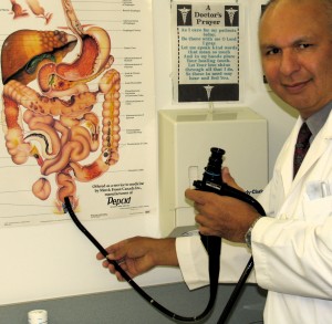
Dr. Bharwani demonstrating the use of flexible sigmoidoscope - checks left side of the colon for average risk patient.
Dr. Bharwani demonstrating the use of flexible sigmoidoscope – checks left side of the colon for average risk patient.

Rightly so, the media immediately picked up the conclusions of a recent study (Colonoscopic Polypectomy and Long-Term Prevention of Colorectal-Cancer Deaths) published in the New England Journal of Medicine (February 23, 2012).

Among 2602 patients who had adenomas (pre-malignant polyps) removed during participation in the study, after a median of 15.8 years, had 53 per cent reduction in death from colorectal cancer.

This indeed is an important conclusion. As New York Times (February 22/12) said, “Although many people have assumed that colonoscopy must save lives because it is so often recommended, strong evidence has been lacking until now.” This study has some limitations in that it was not a double blind or randomized trial.

The Times article also says, “The new study did not compare colonoscopy with other ways of screening for colorectal cancer and so does not fully resolve a longstanding medical debate about which method is best. Tests other than colonoscopy look for blood in the stool or use different techniques to examine the intestine. All the tests are unpleasant, and people are often reluctant to have them.”

In fact, a study from Spain found that when people were offered a stool test, only 34.2 per cent took it. The figure for colonoscopy was even worse: 24.6 per cent.

One thing most physicians agree is that it is important to get some type of screening test for colorectal cancer starting at age 50. Research indicates that not every polyp turns into cancer, but that nearly every colorectal tumor starts out as an adenomatous polyp. In the general population this type of polyp is found in about 15 per cent of women and 25 per cent of men.

So, not every 50 year old and older is at risk of having colorectal cancer. Some are at average risk, some are at moderate risk and some are at high risk. It all depends on your personal and family history.

Not all doctors who do colonoscopies are good at finding polyps. Studies have shown that polyps in the right side of the colon are more often missed than on the left side.

Good news is, colonoscopy does not have to be done every year. If there are no polyps, it is recommended just once every 10 years. People with polyps are usually told to have the test every three to five years depending on the size and kind of polyp.

Colonoscopy should be used judiciously. It is invasive and expensive. It carries small risks of bleeding or perforation of the intestine. It requires sedation, a day off work, and patients must take strong, foul-tasting laxatives to clean out the intestines.

Dr. Winawer, one of the authors of the new study is quoted in the Times article saying, “Any screening is better than none. The best test is the one that gets done, and that gets done well.”

So, what are you waiting for. Talk to your doctor about your risk (average, moderate, high), your options, the advantages and disadvantages of each test and go for it. It may save your life.

Honey, where is my bottle of laxative…I said laxative not a bottle of wine.

Start reading the preview of my book A Doctor's Journey for free on Amazon. Available on Kindle for $2.99!

Anal Cancer Can be Misdiagnosed as Hemorrhoids

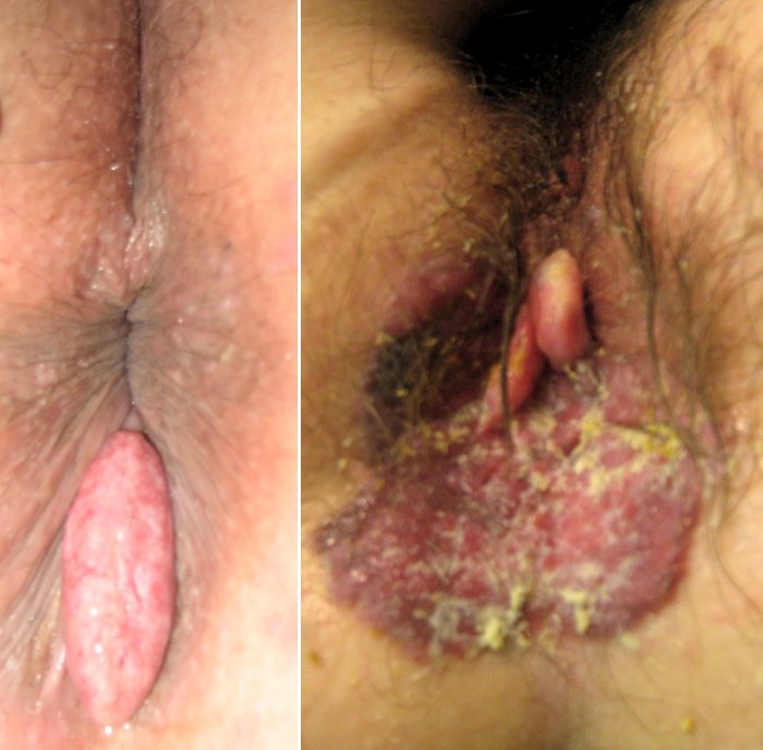
Anal Cancer

You may recall, Farrah Fawcett, one of the Charlie’s Angels, was diagnosed with anal cancer in 2006. Three years later she died.

There is no need to panic. Anal cancer is fairly uncommon. It accounts for about one to two per cent of gastrointestinal cancers. About 4,000 new cases of anal cancer are diagnosed each year in the U.S.A., about half in women. Approximately 600 people will die of the disease each year.

In Canada, incidence of anal canal tumours is approximately 515 cases per year with annual incidence rate of 1.3 per 100,000 population. Review of cancer registry by researchers has shown that the incidence of anal cancer in Canada is increasing.

Anal cancers can be just outside the anus (perianal) or inside the anus. The anal canal extends from the anal verge to the upper border of the anal sphincters, and is approximately four to five cm in length. The skin for a five cm radius around the anal verge is called the perianal skin or anal margin.

What are the risk factors for developing anal cancer?

We do not know the exact cause of most anal cancers. But we know certain risk factors are linked to anal cancer. Most people with anal cancer are over 50 years old. Having anal warts significantly increases the risk. Anal warts are caused by infection with the human papilloma virus (HPV).

Persons who participate in anal sex are at an increased risk. Use of condoms is highly recommended to reduce the risk. Harmful chemicals from smoking increase the risk as well. People with weakened immune systems, such as transplant patients who must take drugs to suppress their immune systems and patients with HIV (human immunodeficiency virus) infection, are at a somewhat higher risk.

People with long-standing anal fistulas or open wounds are at a slightly higher risk. People who have had pelvic radiation therapy for rectal, prostate, bladder or cervical cancer are at an increased risk.

What are the symptoms of anal cancer?

Mostly they are no different than symptoms of hemorrhoids. That is why patients should stop saying, “Doctor, my hemorrhoids acting up again.” When you see your doctor, say what symptoms you have and let him/her make the diagnosis.

Most patients will complain about bleeding, itching, feeling of a lump, may have pain, narrowing of stools, discharge and staining of underwear and in advanced cases there may be enlarged groin lymph glands.

Biopsy is required to confirm the diagnosis. Treatment of anal cancer depends on the extent of the problem and may include surgery, radiotherapy and chemotherapy. Anal cancer can be prevented or picked up in early stages by eliminating the risk factors mentioned earlier and having your butt checked out on a regular basis. Follow the protocol for screening for anal, rectal and colon cancer. For more information, visit my website: www.nbharwani.com.

Start reading the preview of my book A Doctor's Journey for free on Amazon. Available on Kindle for $2.99!