New Guidelines for Use of Testestorone in Male Menopause

Do men undergo menopause? If yes, then how do we diagnose it and treat it?

The medical term for male menopause is andropause. “Andro” stands for androgen – a male sex hormone, such as testosterone or androsterone, which controls the development and maintenance of masculine characteristics. Andropause is also known as ADAM (androgen decline in the aging male).

Amongst physicians, some believe in male menopause and others do not. There is not a consensus case definition for androgen deficiency. The main reason is that the clinical manifestations of testosterone deficiency are usually subtle and variable. This results in poor understanding of the condition.

In order to provide an evidence-based foundation for diagnosis and management of andropause, the Endocrine Society recently published clinical practice guideline (J. Clin. Endo. Metab. 2010; 95:2536-59) to help physicians treat this poorly understood condition.

The full title of the document is, “Testosterone Therapy in Men With Androgen Deficiency Syndromes: An Endocrine Society Clinical Practice Guideline.”

At what age does testosterone deficiency in men start?

The precise age at which testosterone levels start to decrease is not known. Testosterone levels decline by one to two per cent per year in older men, and the circadian (biological rhythm) variability of levels present in younger men is also commonly lost with aging, says the guideline. Hence, the symptoms of testosterone deficiency in men vary with the age of onset and degree of testosterone deficiency.

Aside from the normal aging decline in testosterone production by the testicles, there are many other reasons why testicular function may fail. Such as: testicular injury, infection, tumours, surgery and effect of other hormonal problems.

What kind of symptoms testosterone deficiency produces?

According to the guideline, the signs and symptoms most consistent with testosterone deficiency include decreased libido, erectile dysfunction, gynecomastia (enlargement of male breast), loss of body hair, hot flushes/sweats, bone loss and/or low-impact fractures, absence of sperms in the semen/infertility, and incomplete sexual development.

A variety of less-specific symptoms also may be attributable to testosterone deficiency: decreased energy or mood, sleep disturbance, poor concentration, modest anaemia and increased body fat with decreased muscle bulk/mass, says the guideline.

How to make a diagnosis?

Besides evaluating the clinical symptoms, a morning total testosterone level is the recommended initial test for androgen deficiency. If low, then this should be repeated to confirm the results. Some patients who suffer from chronic illnesses might require measurement of free testosterone levels.

Testosterone level is highest in early morning and can decrease by 35 percent in the mid-afternoon and evening. Early morning testosterone level less that 7 nmol/l indicates that a man has poor testicular function. This will warrant further investigation to find the reason for low level. Is it a primary testicular problem or secondary to other medical conditions?

The guideline is very specific in saying that testosterone deficiency should not be diagnosed without the presence of both symptoms and low testosterone levels.

Does testosterone therapy work?

The guideline says that the randomized trials of testosterone replacement in men with testosterone deficiency have shown consistent improvement in bone density, lean body mass with concomitant reduction in fat mass and sense of physical well-being.

The trials were less consistent in effects on muscle strength, libido, erectile function, quality of life, depression, cognition and muscle strength. Testosterone replacement has not been demonstrated to reduce fractures. Many of the trials are limited by small sample size and short follow-up.

Testosterone treatment is not recommended in men with breast or prostate cancer, elevated PSA, and/or unevaluated prostate abnormality, those at high risk for prostate cancer, those with severe lower urinary tract symptoms or in men with hematocrit greater than 50 per cent, untreated sleep apnea or poorly controlled heart failure. Men treated with testosterone replacement should be evaluated on regular basis.

Start reading the preview of my book A Doctor's Journey for free on Amazon. Available on Kindle for $2.99!
Snowbirds – Be Aware of the Risks of Blood Clots in the Legs

Fall is here. Winter will be here soon. Canadian snowbirds are heading south. This involves long hours travelling by road or by plane. Studies have shown a two- to four-fold increased risk of blood clots in the legs following air travel. Similar risk applies to travel by road. The condition is called venous thrombo-embolism (VTE).

There are several risk factors. A population-based case-control study of adults receiving treatment for their first VTE found that long-distance travel (≥4 hours) doubled the risk of VTE. The effect was greatest in the first week after travel but remained elevated for 2 months. Travel by air increased the risk to the same extent as travel by bus, train, or car, suggesting that the increased risk of air travel is due primarily to prolonged immobility.

Women who used oral contraceptives, travellers who were overweight with BMI >30 kg/m2, and those with height >1.9 m (approximately 6 ft 3 in) were at increased risk. People shorter than 1.6 m (approximately 5 ft 3 in) had an increased risk of VTE only after prolonged air travel. Cancer, dehydration and smoking cigarettes also increase the risk of VTE.

A blood clot in a superficial vein is known as superficial thrombophlebitis. This condition is usually not serious or life threatening.

A blood clot in a deep vein of a leg is known as deep vein thrombosis (DVT). This is a serious condition as the clot may dislodge, travel through the blood stream and plug a vessel in the lung (pulmonary embolism). Pulmonary embolism (PE) can be fatal.

The calf muscles act as a second pump (first pump being your heart). The contraction of the calf muscles and the valves in the deep veins help push the blood from the lower extremity towards the heart.

DVT occurs when the blood moves through deep veins in the legs more slowly than normal or when there is some condition that makes blood more likely to clot. Two common examples are: when you are bedridden (after surgery, injury or chronic illness) or when you sit still for a long time (such as during a long plane flight or a long road trip). Under these conditions the blood moves more slowly and stagnation promotes clotting.

Symptoms of DVT include swelling, redness, pain, or tenderness, and increased warmth over the skin. It may be difficult to distinguish from muscle strain, injury, or skin infection. Diagnosis is confirmed by special radiological tests. Symptoms of PE range from mild and nonspecific to acute, resembling heart attack or stroke. Once a clot has travelled to the lungs, common symptoms of PE are chest pain and shortness of breath.

There are measures you can take to prevent VTE. All travellers should keep hydrated with non-alcoholic beverages, wear loose-fitting clothing, do frequent calf muscle contraction and make efforts to walk and stretch at regular intervals during long-distance travel. Compression stockings may be beneficial to travellers with other risk factors for VTE. Currently no convincing data suggest that pharmacologic interventions reduce the risk of significant VTE during travel.

Treatment of VTE is with blood thinners (anticoagulants) like heparin and Warfarin to prevent pulmonary embolism. The blood thinners do not dissolve the clot. They stop the clot from getting bigger, prevent the clot from breaking off and reduce the chances of having another blood clot.

The body takes its own time to dissolve the clot or the clot may get organized and form scar tissue, permanently blocking the vein or damaging the valves. Normally, no tests are done to check if the clot is still present as the tests can be inconclusive or confusing.

Start reading the preview of my book A Doctor's Journey for free on Amazon. Available on Kindle for $2.99!
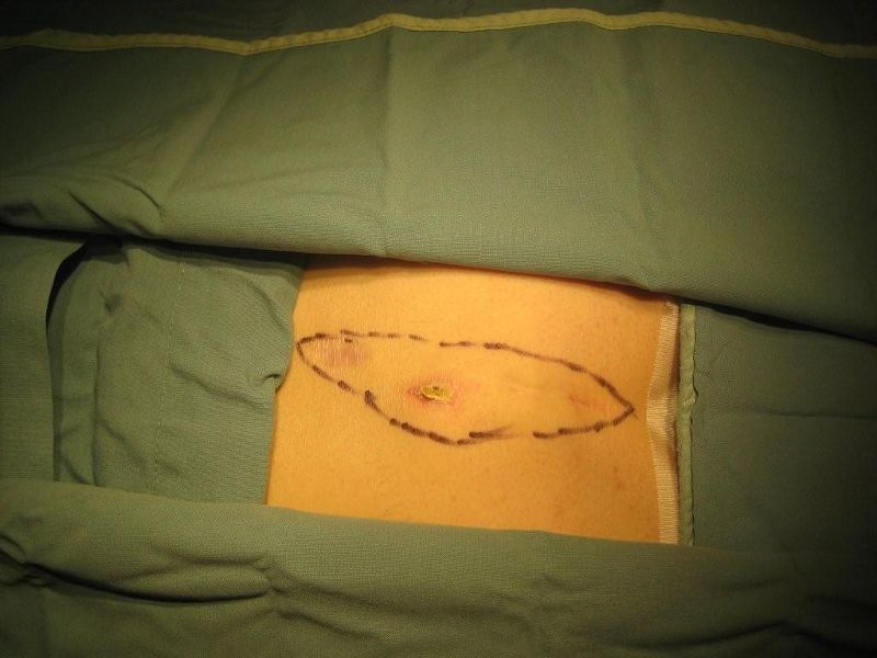
Case #1 – Pilonidal Disease

The pilonidal disease most commonly occurs between the buttocks, close to the tailbone. This condition has been described since 1847.

The term pilonidal means hair-nest.

It can also occur in other areas such as beard, the armpit, the belly button and the web spaces of the hands (in barbers) and feet.

The condition results from a reaction to hairs embedded in the skin, commonly occurring in the cleft between the buttocks. The disease is more common in men than women and frequently occurs between puberty and age 40. It is also common in obese people and those with thick, stiff body hair.

Pilonidal disease is a spectrum of three conditions:
-acute pilonidal abscess,
-chronic pilonidal abscess or sinus,
-unhealed pilonidal surgical wound.

For many years, experts believed that this was a congenital condition. In 1946, Patty and Scarff challenged this theory and drew attention to the role of hair in the origin of this problem. Current evidence strongly indicates an acquired origin for pilonidal disease, with most infections being related to penetration of the skin by hair through small midline pits.

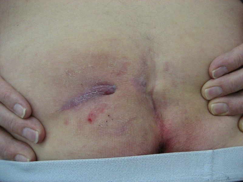
Example of pits and infection:
Example of pits and infection.

Where do these pits come from? Some people believe they are congenital while others believe them to be enlargement of hair follicles. These pits have sinus openings through which the hairs penetrate. Hirsutism in the buttock and perineal area appears to be associated with the development of pilonidal disease.

Management of the condition depends on the type of presentation.

Acute pilonidal abscess needs to be drained immediately once the diagnosis is made. The area should be kept shaved. Daily bath or shower will keep the area clean. Once healed, it may become necessary to excise the midline pits under local or general anaesthesia to prevent recurrence.

Treatment of chronic pilonidal abscess or sinus remains controversial as no one treatment has proved superior. The choices are:
-non-operative treatment with repeated phenol injections
-conservative excision of the sinus openings and midline pits
-laying open the sinus tract and stitch the skin margins to fibrous tissue (marsupialization)
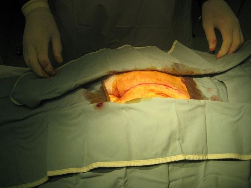
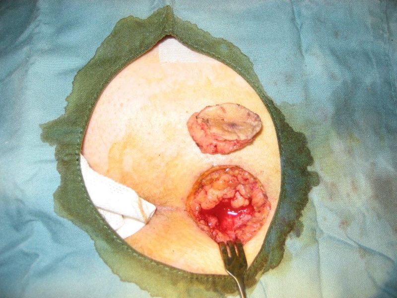
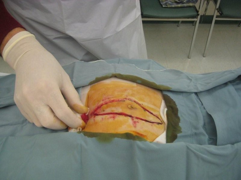
-wide excision with or without different types of closures of the skin.

Example of wide excision:
Example of wide excision

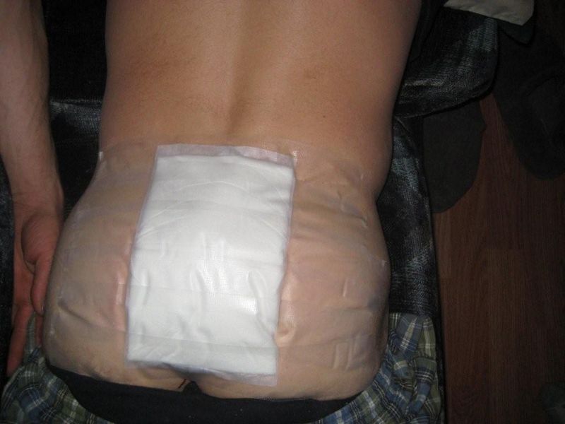
The unhealed pilonidal surgical wound and recurrence of pilonidal disease after initial treatment is very common. Management of this problem can be very difficult. To start with, the unhealed wound should be curettaged to control the excessive granulation tissue (healing tissue which fills the wound), and the surrounding skin should be shaved weekly. The wound should be kept clean and dry with gauze. Strapping the buttocks apart may help prevent the continuous shearing movement during walking.

The wound may take six to eight weeks to heal. Quite often healing does not occur. Then a skin graft or some form of plastic flap may help the healing process.

If the wound can be closed, it will need to be kept clean and dry until the skin is completely healed.

If the wound must be left open, dressings or packing will be needed to help remove secretions and to allow the wound to heal from the bottom up.

After healing, the skin in the buttocks crease must be kept clean and free of hair. This is accomplished by shaving or using a hair removal agent every two or three weeks until age 30. After age 30, the hair shaft thins, becomes softer and the buttock cleft becomes less deep.

Overall, treating pilonidal disease should not be taken lightly as the results may be disappointing. There is no one good treatment which works for all patients with pilonidal disease

Pilonidal disease image 1
Pilonidal disease image 2
Pilonidal disease image 3
Pilonidal disease image 4
Pilonidal disease image 5

For more information visit:
-Another article on my website – Pilonidal Cyst
American Society of Colon and Rectal Surgery
Pilonidal disease support group

Start reading the preview of my book A Doctor's Journey for free on Amazon. Available on Kindle for $2.99!
The Role of Exercise in Some Intestinal Disorders

In 2001, Gut – An International Journal of Gastroenterology and Hepatology, published a review article from the Netherlands titled “Potential benefits and hazards of physical activity and exercise on the gastrointestinal tract.”

It is an interesting review. It says that acute strenuous exercise may provoke gastrointestinal symptoms such as nausea, heartburn, diarrhea and gastrointestinal bleeding. This happens especially during vigorous sports such as long distance running and triathlons. About 20 – 50 per cent of endurance athletes are hampered by these symptoms. This may deter them from participation in training and competitive events. But it has no long term effect on their health.

In contrast, the article says, repetitive exercise periods at a relatively low intensity may have protective effects on the gastrointestinal tract.

There is strong evidence that physical activity reduces the risk of colon cancer by up to 50 per cent. The primary postulated mechanism, according to the article, is that physical activity reduces intestinal transit time which would limit the time of contact between the colon mucosa and cancer promoting contents.

Several studies have been published on the relationship between physical activity and gall stones. Still more work needs to be done. Regular exercise may reduce the chance of developing gall stones.

A limited number of studies have investigated the preventive effect of physical activity on inflammatory bowel disease (Crohn’s disease and ulcerative colitis). The article says, “While the preventive effect of physical activity remains inconclusive, it has become clear that physical activity is not harmful for patients with inflammatory bowel disease….”

Physical activity in patients with inflammatory bowel disease should be encouraged as these patients have muscle weakness and are at risk of osteoporosis, especially if they are on steroids for the treatment of their disease.  Exercise will improve physical health, general well being, perceived stress and quality of life.

A possible role of physical activity in reducing the risk of diverticular disease has been reported in the medical literature. It has been suggested that diverticular disease was more prevalent among people with sedentary occupations than in more active occupations. An increase in colonic motor activity via hormonal, vascular, and mechanical aspects, leading to a reduction in colonic transit time, was postulated as an underlying mechanism.

Regular physical activity and exercise has shown a positive effect on reducing constipation. The review article mentions two case control studies that showed the defecation pattern of runners was “better” (less firm stools, higher defecation frequency, higher stool weight) than in inactive controls. Further research is required to confirm these findings.

In conclusion, strenuous exercise may induce gastrointestinal symptoms such as heartburn or diarrhoea, which may deter people from participating in physical activity. These symptoms are usually transient.

Physical activity, mostly performed at a relatively low intensity, may also have protective effects on the gastrointestinal tract. There is strong evidence that physical activity reduces the risk of colon cancer. Less convincing evidence is found for gall stones and constipation.

Physical activity may reduce the risk of diverticulosis, gastrointestinal bleeding, and inflammatory bowel disease, although up to now there has been little research to substantiate this. Physical activity does not interfere with the healing process in inflammatory bowel disease and will probably not reduce the risk of rectal and gastric cancer, says the article.

Start reading the preview of my book A Doctor's Journey for free on Amazon. Available on Kindle for $2.99!