Trip to Jamaica! – Evidence Based Medicine

You see a doctor for a medical problem. He advises you to follow certain treatment. Do you ever ask him: Doctor, is there any scientific evidence to show that this treatment works?

Most of us trust our doctor and are too polite to ask him such a question. Instead we rely on our neighbors, friends and families to give us a second and a third opinion.

Those who are computer literate surf the internet. But you know what happens there – there are thousands of references to search for an answer.

What do doctors do when they are looking for best evidence in their practice?

Doctors go back to their text books, read medical journals, talk to their colleagues, have case conferences in hospital or attend medical meetings at exotic places.

My surgical associate, Dr. Brzezinski and I just got back from Montego Bay, Jamaica where we attended a two day conference on Evidence Based Medicine in Gastroenterology.

It was a very interesting conference. The location was beautiful – an ideal environment to learn something! The warm ocean breeze, carrying important scientific knowledge, penetrates your brain without difficulty!

There were experts from Europe, Canada and Jamaica. Main discussion was on the problems of the esophagus and stomach.

As I have said many times here, medicine is an imperfect science. Quite often the practice of medicine is an art than science. And the discussion at the Jamaican conference again confirmed that.

Only about 10 to 20 percent of what we do in medicine is evidence based. That means it is scientifically proven. The rest is based on what each one of us think is correct, what we think is best for a particular patient, it is economical and safe.

The advantage of evidence based medicine is that it helps optimize patient care and minimize variation in best practice.

The problem is that in most cases there is not enough evidence available. The clinical decision making is a very complex process because no two patients respond to a treatment in exactly the same manner.

Therefore, evidence based medicine in clinical practice is quite often not relevant.

But in spite of imperfections in medical practice, we continue to treat hundreds and thousands of patients each day. Most of them do well and respond to treatment.

Some get better just by talking to a sympathetic doctor.

Some get better by taking an aspirin and going to bed.

Some get better by doing nothing – may be a shot of brandy. Or Jamaican style – don’t worry, be happy.

Some get better by following the principles of ELMOSS – exercise, laughter, meditation, organic/healthy food, stress relief, and by giving up smoking.

But eventually, most people do get better. Time is a good healer – unless you are suffering from an incurable disease.

So, medicine is not a rocket science. But you have to know the human anatomy, physiology, pharmacology, and pathology. Then you have to put all this knowledge together and pass few exams. Then you can call yourself a doctor of medicine and surgery!

Isn’t that easy? It just takes 10 to 15 years of your life. Then you start practice and find out that only 20 percent of what you practice is based on pure science! But you can say – I have been to Jamaica!

Seriously – next time you are sick………………well see your doctor first!

Start reading the preview of my book A Doctor's Journey for free on Amazon. Available on Kindle for $2.99!
Breast Feeding

Human milk is the ultimate form of early nutrition for children, and the search for the ideal substitution infant formula will never be concluded satisfactorily.

Dr.Yap-Seng Chong, BMJ September 20, 2003

The year 2003 marks the 12th annual World Breastfeeding Week. It is celebrated on the 40th week of the year (October 1-7) because from conception to birth, breast feeding is initiated on the 40th week.

One would think that breast feeding would be a natural sequential process after pregnancy and birth. Body’s hormonal system is designed in such a way that the breasts are ready with milk when the baby arrives.

Then why have World Breastfeeding Week?

A report shows that in Canada, the overall rate of breast feeding initiation was 75 percent in 1991 and 1992. Fifty-four percent of women were still breast feeding at three months and 30 percent at six months of age.

In 1995, 60 percent of women in the United States were breastfeeding either exclusively or in combination with formula feeding at the time of hospital discharge; only 22 percent of mothers were nursing at six months, and many of these were supplementing with formula, says another report.

The target is to have more than 75 percent of mothers breastfeed their babies in the early postpartum period and to have at least 50 percent to continue breastfeeding until their babies are six months old

What are the obstacles to the initiation and continuation of breastfeeding?

There are many. These include physician apathy and misinformation, insufficient prenatal breastfeeding education, disruptive hospital policies, inappropriate interruption of breastfeeding, early hospital discharge, lack of timely routine follow-up care and postpartum home health visits.

Other obstacles are: mother’s place of employment (especially in the absence of workplace facilities and support for breastfeeding), lack of broad societal support, media portrayal of bottle-feeding as normative, and commercial promotion of infant formula through distribution of hospital discharge packs, coupons for free or discounted formula, and television and general magazine advertising.

These obstacles will have to be removed to encourage young mothers to provide the best possible care for their infants.

Extensive research has shown compelling advantages to infants, mothers, families, and society from breastfeeding and the use of human milk for infant feeding. These include health, nutritional, immunologic, developmental, psychological, social, economic, and environmental benefits.

Both the American Academy of Pediatrics and the Canadian Pediatric Society have recommended breast feeding as the preferred mode of infant feeding.

The World Health Organization and UNICEF have developed explicit guidelines to encourage breast feeding around the world.

More information can be obtained by visiting the websites of these organizations. Locally, you can phone Community Health Services (403-502-8200) and get more information and help.

All communities worldwide need to protect, promote and support breastfeeding. And to remove barriers which inhibit young mothers to feed their infants on demand. We should encourage mothers to continue breastfeeding for at least six months.

Start reading the preview of my book A Doctor's Journey for free on Amazon. Available on Kindle for $2.99!

Breast Lump Evaluation

A breast lump in a woman or a man is an important clinical finding – never to be ignored. It is also a source of anxiety for the patient and his or her family.

The usual questions are: What is it doctor and what investigations are required to check for cancer?

Investigations of a breast lump starts with a history and physical examination. Physical examination is best done between seven to 10 days from the first day of the menstrual cycle.

The clinical history will establish how long the lump has been present. Whether there has been any change. And if there is a previous history of breast biopsy or breast cancer.

Risk factors for breast cancer will be noted (patient’s age and family history). Absence of risk factors does not decrease the probability of cancer.

Examination of the breasts will confirm the presence and establish the character of the lump. Hard, irregular, tethered or fixed, or painless lump may be suggestive of cancer. Axillary (armpit) and neck areas are checked for lymph glands. The predictive value of physical examination (to say whether the lump is benign or malignant) in experienced hands is about 75 percent.

Mammograms in younger women are not very helpful. But after mid-30s the value of mammography increases. Quite often mammography can clarify the nature of the lump. It can also detect non-palpable lumps.

The overall level of sensitivity of mammography in palpable breast cancers may be no more than 82 percent. It may be even lower in premenopausal women. A negative mammogram in the presence of a persistent lump does not exclude malignancy.

Fine-needle aspiration biopsy done in the office can establish whether a breast lump is solid or cystic. When the lump is solid, cells can be aspirated for examination under a microscope. If fluid is obtained during aspiration and the lump disappears then it is cystic.

In a cystic lump, if the fluid is not bloody then no specimen needs to be sent for examination and no further investigation is necessary. Bloody fluid may be suggestive of cancer and should be sent for microscopic examination.

Fine-needle aspiration biopsy should provide satisfactory specimen for microscopic examination in 90 percent of the cases with an accuracy rate of 95 percent to detect presence of cancer cells.

When physical examination, mammography, and microscopic examination of aspirated specimen indicate cancer then the probability of this being confirmed by surgery is more than 99 percent. If all three tests are negative then the lump being cancerous is less than one percent.

Core-biopsy, where a bigger sample (a core of tissue) is taken from a breast lump, has 90 percent predictive value for breast cancer. A negative biopsy may make it unnecessary to do an open surgical biopsy.

Ultrasound of the breast lump is an alternative method to distinguish a cyst from a solid lump. Ultrasound is useful when a non-palpable lump is seen on a mammogram.

Nuclear medicine techniques (Miraluma scan) have been used to evaluate breast cancer. Its predictive value for cancer is around 80 percent for a palpable lesion and 50 to 70 percent for a non-palpable breast lump found on mammography.

Whenever reasonable doubt remains about cancer (since none of the tests have a one hundred percent accuracy rate), a surgical open biopsy is undertaken.

Only about one in every five to 10 breast biopsies leads to a diagnosis of cancer. But every breast lump should be investigated thoroughly.

Start reading the preview of my book A Doctor's Journey for free on Amazon. Available on Kindle for $2.99!

Rectal Pain (Proctalgia Fugax)

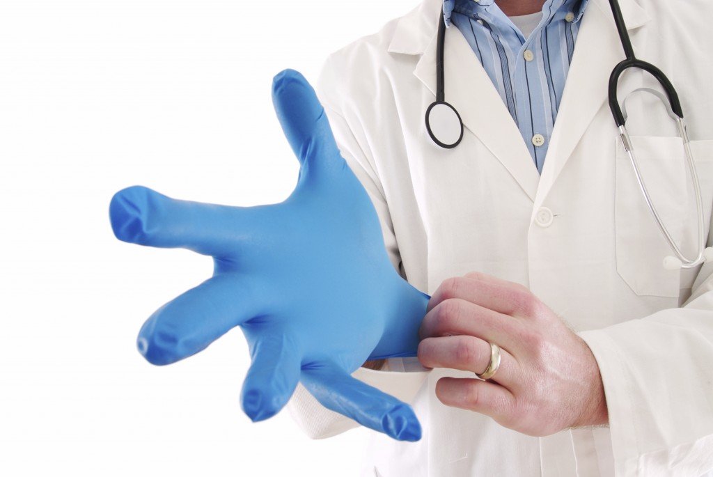
A doctor putting on his glove. (iStockphoto/Thinkstock)

A patient asks: I have pain in my rectal area and I have been told that I have proctalgia fugax. What is this condition and how can I get some relief?

There are at least six common causes for rectal and anal pain: pruritus (itch), external thrombosed hemorrhoid (a blood clot), prolapsed internal thrombosed hemorrhoids, fissure (tear), abscess, and fistula (tunnel).

Proctalgia fugax is not part of the list as it is not that common. But unfortunately it is not that rare either. It occurs in about 14 percent of healthy people. Seventy five percent of these are women.

Proctalgia fugax falls under the category of “unexplained rectal and anal pain”. Other conditions under this group are levator ani syndrome and coccygodinia.

Let us try and understand some anatomy first.

Colon ends in the pelvis to become sigmoid, rectum and anus. Sigmoid and rectum act as storage area for fecal matter. At a socially convenient place, the anal sphincters (valves) relax to allow us to defecate.

Anal canal is surrounded by two circular muscles known as internal and external sphincters. Rectum is surrounded by and held in place by pelvic floor consisting of a group of muscles called levator ani. Coccyx is the tail end of the spine, not too far from the anal canal.

Proctalgia means pain in the rectum. Fugax means flying, fleeting, momentary like a fugitive – trying to elude justice!

Proctalgia fugax is an intensely painful spasm in the rectal area that begins abruptly and lasts for several minutes. It can begin during sleep, defecation, urination, or intercourse. The character of the pain has been compared to a charley horse.

Sharp cramp or stabbing pain may awaken the patient from sleep. It lasts less than 30 minutes and may radiate to the coccyx or perineum. It may only occur once a year or several times a week. Pain may be severe enough to cause sweating and palpitation. There may be a desire to have a bowel movement, yet pass no stool.

It is thought that a sudden spasm of the levator muscle complex or the sigmoid colon can result in proctalgia fugax.

It is believed that people who frequent the toilet are at greatest risk. Professionals, managers, and perfectionists are more likely to be afflicted. Stress and anxiety plays a role in precipitating the pain.

The diagnosis is based almost entirely on the patient’s symptoms. Clinical examination is usually negative. Patients should undergo flexible sigmoidoscopy to screen for other causes of ano-rectal diseases. Careful pelvic and prostate examinations should be undertaken. Ultrasound or CT scan of the pelvis may be necessary.

Patients with levator ani syndrome experience pain for hours to days. The pain is most often constant or rhythmic and may be likened to sitting on a ball or feeling like a ball (or corncob) was inside the rectum. Pain may be caused by defecation, sexual intercourse, sitting for long periods, and stress or anxiety. The pain is probably due to spasm of the pelvic floor muscles.

Coccygodynia is a cramp or ache in the tailbone and typically results from injury to the coccyx or arthritis. Movement of the coccyx can reproduce the pain.

Pain from proctalgia fugax, levator syndrome, and coccygodynia may be hard to differentiate.

Treatment is often unrewarding. Some of the measures worth trying are: reassurance, hot baths, bowel regimens, message therapy, perineal strengthening exercises, pain killers, anti-inflammatory, muscle relaxants, topical nitrates, tranquillizers, calcium channel blockers, acupuncture, and psychiatric evaluation.

Unfortunately, proctalgia fugax is one of the many medical conditions for which there is no good explanation or treatment.

Start reading the preview of my book A Doctor's Journey for free on Amazon. Available on Kindle for $2.99!